Sunday, March 4, 2012

Laparoscopy in General Surgery


Laparoscopic cholecystectomy


This is a widely used procedure in general surgery. Prior to surgery the procedure is explained to the patient and informed written consent is taken. The appropriate assessment of patient’s fitness for surgery is carried out. This includes investigation of cardiovascular and respiratory system if history suggests these to be risk factors, a full blood count and biochemical profile. Blood coagulation is checked if there is history of jaundice. 
At the operating theatre the patient is positioned in the operating table. The patient is given prophylactic antibiotics at the time of induction. The patient is anaesthetized. And pneumoperitoneum is created. Four ports are placed in the abdomen.
The cystic duct and cystic artery are carefully defined. Then the cystic duct is clipped and divided and the gallbladder is removed from the bed then removed from the body via the umbilicus.
Then the co2 is removed and trocars are withdrawn. The access sites are sutured. Other practices of laparoscopy in General surgery.


Diagnostic Laparoscopy


Appendectomy
The surgeon typically stands on the left of the patient, and the assistant stands on the right. The standard approach is to place 3 trocars during the procedure. Two of these have a fixed position ( umbilical, suprapubic); the position of the third, which is placed in the right periumbilical region, may vary greatly depending on the patient's anatomy. After gaining access the whole abdomen is visualized and the appendix is identified.

Hernia Repair.
The port placement differ according to the site of the hernia


Spleenectomy


Anti-Reflux Procedures
The most common anti-reflux surgical procedure performed for treatment of GORD is a Nissen fundoplication . The fundus of the stomach is wrapped around the lower portion of the esophagus and anchored securely below the diaphragm. If there is a hiatal hernia, the hernia will also be fixed using a laparoscope and five small incisions


Paraesophageal Hernia Repair


Gastric Procedures
Since the advent of H2-receptor blockers and proton pump inhibitors elective surgery for ulcer disease is unusual, but in emergency surgery for complications such as perforation and gastric outlet obstruction, laparoscopic procedures are appropriate. These procedures include primary repair with omental patch for perforation and gastrojejunostomy bypass in cases of obstruction. Management of bleeding by laparoscopic techniques is not being done but Laparoscopic gastrectomy for benign and malignant gastric tumors has been done


Small Bowel Procedures


Colorectal Surgery


Laparoscopic Procedure


 Gaining Access
Access to the peritoneal cavity and creation of pneumoperitoneum is the initial step in laparoscopy.There are different methods of primary access in laparoscopy. The popular ones being the Veress needle technique and Hasson’s technique. Veress needle technique still being used by many surgeons and gynaecologists is regarded by them as the Gold standard. As the veress needle and the first trocar afterward are introduced blindly, this method is called closed method contrary to the open technique named as Hasson’s technique where peritoneum is approached by open dissection of tissues.

The veress needle technique
Umblicus is the site chosen as the primary access site. The reasons are it is in the midline and it is naturally a weak area due to absence of all the layers.
The anterior abdominal wall should be lifted or stabilized before the insertion of the needle or stabilized before the Veress needle is inserted. Only three attempts for successful pneumoperitoneum establishment are acceptable, fourth attempt should be made in an alternative site. One of these sites is the Palmer’s point localized about 3 cm below the left costal margin in the midclavicular line. This is the site especially recommended in extremely obese and thin patients,
The Veres needle should be inserted in angle from 45° in non-obese to 90° in obese patients. There are various methods like hanging drop of saline test, the “hiss” sound test, aspiration and syringe test, that prove correct localization of the needle.
Then pneumoperitoneum is created using CO2 insufflator.
The last is the introduction of the first trocar, it has a potential in damaging major vessels or organs.So it is essential to elevate the abdominal wall.It should be introduced carefully with the rotatory motion. Just after the insertion of the first trocar, the insufflation cord should be connected to maintain the pneumoperitoneum.

Secondary Access
After taking the primary access secondary access of the other trocars should take place. The site and no of placement differ according to the procedure. Telescope should be in the middle of instruments. Manipulation angle of instruments usually 60 degree but can differ according to procedure. The picture shows the placement of camera, target and instrument ports.

When inserting the ports, abdominal wall has to be pulled up and trocar inserted in a twisting manner. This has to be stopped when the trocar enters the cavity.

History of Laparoscopic Surgery


1902Georg Kelling, of Dresden, Saxony, performed the first laparoscopic procedure in dogs. He used a cystoscope to peer into the abdomen of the dog after first insufflating it with air. He also used filtered atmospheric air to create a pneumoperitoneum, with the goal of stopping intra abdominal bleeding.

1910- Hans Christian Jacobaeus of Sweden reported the first laparoscopic operation in humans. This was done in USA. The instrument used was a proctoscope of a half inch diameter and ordinary light for illumination. The procedure was mainly inspecting abdominal cavity. This was called “organoscopy”.
An Early Laparoscopic Surgery

An early Lparoscopic Surgery


1911- H.B.Jacobaeus, again coined the term “laparothorakoscopie” after using this procedure on the thorax and abdomen. Heused introduced the trocar inside the body cavity directly without employing a pneumoperitoneum.

1920 -Zollikofer of Switzerland discovered the benefit of CO2 gas to use for insufflations, rather than filtered atmospheric air or nitrogen.

1929 - Heinz Kalk, a German gastroenterologist who is considered the founder of the German School of Laparoscopy developed a specific lens system and a dual trocar approach. He used laparoscopy as a diagnostic method for liver and gallbladder disease.

1934 - John C. Ruddock, M.D., F.A.C.P., pioneer in laparoscopy, an American internist described laparoscopic as a good diagnostic method, many times, superior than laparotomy. 


1936- Boesch ot Switzerland is credited for doing the first laparoscopic tubal sterilization.

1938 - J Veress, of Hungary, developed the spring-loaded needle. It main purpose was to perform therapeutic pneumothorax to treat patients suffering from tuberculosis. It current modifications make the “Veress” needle a perfect tool to achieve pneumoperitoneum during laparoscopic surgery.
 
1944 - Raoul Palmer, performed gynecological examinations using laparoscopy and placing the patients in the Trendelemburg position, so air could fill the pelvis. He also stressed the importance of continuous intra-abdominal pressure monitoring during a laparoscopic procedure. 
1950- The first publication on diagnostic laparoscopy by Raoul Palmer.

1953-The rigid lens system was discovered by Professor Hopkins. By making this instrument he has revolutionized the concept of laporoscopic surgery.

1960-Kurt semm a German gynaecologist, has invented the automatic insuffator. In late 60 s this was developed into a method which can measure the intra abdominal pressure. This allowed safer laporoscopy.

1970-Laporascopy was widely used by the gynaecologist but general surgeons remained confined to the open surgery.

1972-H.Countay Clarke first time showed laparoscopic suturing technique foe haemostasis.
1980 - Patrick Steptoe, from England started to perform laparoscopic procedures in the operating room under sterile conditions.
1982 - First solid state camera was introduced. This is the start of "video-laparoscopy"

1983- First appendicectomy by Semm, a German gynaecologist.

1985- First laparoscopic cholecystectomy.

1987- First laparoscopic repair of inguinal hernia.

1987- First laparoscopic cholecystectomy using video technique. This procedure has revolutionized the general surgery.

1988- First laparoscopic lymphadenectomy.

1994 - A robotic arm was designed to hold the laparoscope camera and instruments with the goal of improving safety, reducing resource utilization and improving efficiency and versatility for the surgeon.

1996 - First live broadcast of laparoscopic surgery via the Internet.

1997 - Reconnection of the fallopian tubes operation was performed successfully in Cleveland.

1998 - Dr. Friedrich-Wilhelm Mohr using the Da Vinci surgical robot performed the first robotically assisted heart bypass at the Leipzig Heart Centre in Germany.

2001 - Prof. Marescaux used the “Zeus” robot to perform a cholecystectomy on a pig in Strasbourg, France while in New York.
In September 2001, Dr. Michel Gagner used the Zeus robotic system to perform a cholecystectomy on a woman in Strasbourg, France while in New York.
The first unmanned robotic surgery took place in May 2006 in Italy.

Robot Technology in Laparoscopic Surgery.

Introduction


Laparoscopic surgery which has a history of over 100 years is widely used today for pelvic and abdominal surgery. In this minimally invasive procedure the cavity is visualized via a camera inserted in to the body cavity.

 The first laparoscopic surgery has taken place early in the last century and it was done on a dog. It was about decade later the first performance on a human was carried out. First laparoscopic surgery was done without inflating any air into the abdominal cavity. Later filtered atmospheric air was used for this and in 1920 s co2 was first used. At the beginning the abdominal cavity was visualized directly with lenses. Then various lens systems were introduced. However video laparoscopy came into practice in 1980s. With the widespread application of the compact cameras, both laparoscopist and assistants could simultaneously view the operative field on a video screen. By the end of the decade, video-laparoscopy had become standard and operative laparoscopy became widely accepted as a safe and effective surgical approach. A major step forward in the development of laparoscopy was the development of a safer laparoscopic lighting system in the 1950s. Up until that time, intra-abdominal light was produced by a small electric light bulb at the distal tip of the laparoscope. The development of this technology shows the great thinking pattern of human being.

These were widely used in Gynaecology decades before it was used in general surgery. During the last 35 years, gynecologic laparoscopy has evolved from a limited surgical procedure used only for diagnosis and tubal ligations to a major surgical tool used to treat many gynecologic conditions. For many procedures, such as removal of an ectopic pregnancy, treatment of endometriosis, or ovarian cystectomy, laparoscopy has become the treatment of choice.

The use of laparoscopy in general surgery widely came into practice in last 3 decades. But since then it has become versatile. Today it is used for diagnostic and therapeutic purposes in general surgery.

The procedure itself draws interest as it needs special skill and great practice than the open surgery. The basic principle of laparoscopy is simple. A working space within the abdominal cavity is initially established. This is usually accomplished by insufflating the peritoneal cavity with carbon dioxide to a pressure of 10 to 15 mm Hg. The laparoscope is inserted into the abdomen with a trocar and hollow sheath containing a side port for continuous carbon dioxide insufflation, as well as valves and gaskets to allow the insertion and removal of the laparoscope without allowing the carbon dioxide to escape. In an operation, accessory trocars are inserted to introduce laparoscopic instruments to the Abdomen.

The laparoscopic instruments are of two categories. They are equipments for access and exposure and hand instruments for the actual operative procedure. These include the screen, the light source, the insufflater, fibro optic cable, the camera, veress needle, Trocars and other hand instruments needed for the actual procedure. These are graspers, scissors, needle holders and retractors, but all the instruments are long and narrow, and have small jaws that allow the introduction and use of the instruments through trocars. A unique instrument in Laparoscopic surgery is a "clip applicator".

There are a number of advantages to the patient with laparoscopic surgery than an open procedure. They include Reduced hemorrhaging, so the incidence of blood transfusion is less, smaller incision, which reduces pain and shortens recovery time and reduce post-operative scarring. There is less pain so less analgesia is needed; hospital stay is less, and often with a same day discharge. Reduced exposure of internal organs to possible external contaminants thereby reduced risk of acquiring infections.

At the same time there are some risks associated with laparoscopic surgery. They include trauma due to trocar to major blood vessels and major organs and the problems due to co2 insufflation .Also I increased intra-abdominal pressures associated with laparoscopy increase anesthesia-related risks such as aspiration and increased difficulty in ventilation.
 However this technology evolves everyday and becomes more and more versatile. In recent years robot surgery and single incision laparoscopy has been introduced of which the robotic surgery is having the largest impact on clinical care


Laparoscopic Instruments


 Laparoscopic  Tower
Consist of
Ø  High Resolution Monitor-These are flat screen monitors. 

Ø  Light source- A 300-watt xenon light source is usually used. The light is transmitted via flexible, fiber optic bundles connecting the light source to the telescope.

Ø  Viewing Optical System

Ø  Electronic CO2 insufflators-Insuflator (laparoflator) is a device used for introduction of the gas under specified volume and pressure into peritoneal cavity. The CO2 pump should be a high flow insufflation pump with both low flow and high flow settings. Generally, they are capable of delivering high gas flow of 24 L/min, which is limited by the trocar or needle to which it is attached. The insufflation tubing contains a filter that prevents bacterial and viral contamination from possible backflow of surgical smoke, protecting both equipment and health care worker.

·         

Fibro optic cable

This is used to connect light source with optical system. It is necessary to separately check the principles of a precise and accurate treatment with light conductors to avoid damaging thin and sensitive optical glass fibers.

·         Endovideo  Camera


Camera
Laparoscope


Veress needle
 A Veress needle is used to inflate air into the peritoneal cavity. This needle contains a spring-loaded inner sheath. 
This sheath retracts as the needle is advanced through tissue exposing the needle tip. Once the needle enters the peritoneal cavity, and the majority of theresistance on the needle is released, the inner sheath springs forward.  The inner sheath covers the needle tip and protects the intra-abdominal organs from being injured by the sharp end of the needle.


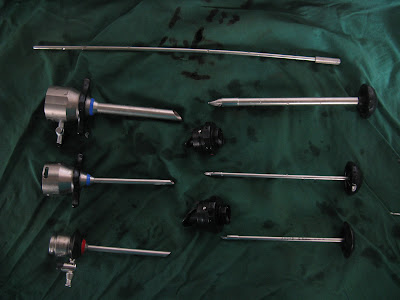
Trocars

The word trocar is originally French meaning three sided. A trocar is a hollow cylinder with an access mechanism on one side which can be a blade (often three-sided), a plastic lip that is used for dilation, or a radial dilating mechanism. On the external side of the trocar is a valve mechanism which allows instruments to be passed in and out of the patient’s insufflated body cavity while maintaining the insufflated space.  Trocars, also known as ports, usually have a valve to which insufflation tubing can be attached to maintain the insufflation pressure.  Most trocars have an outer sheath (also called a cannula or trocar) and an inner obturator, which allows the access.


Hand Instruments in Laparoscopic Surgery

Hand instruments in laparoscopic surgery serve the same basic functions as open surgical instruments - There are graspers, scissors, needle holders and retractors, but all the instruments are long and narrow, and have small jaws that allow the introduction and use of the instruments through trocars. A unique instrument in Laparoscopic surgery is a "clip applicator"


Laparoscopy in Gynaecology.


Laparoscopy and dye test.
This is performed as a diagnostic or a therapeutic procedure in a patient with subfertility. 
In a diagnostic surgery after gaining access to the abdominopelvic cavity via three ports, a colored dye (methylene blue) is injected through the cervix. If the tubes are not blocked the dye should pass along them and spill into the abdomen. In addition to that Laparoscopy allows the internal organs of the abdomen and pelvis to be inspected visually and excludes other problems such as endometriosis, fibroids, ovarian cysts and adhesions.
The procedure becomes therapeutic if adhesiolysis, removal of fibroid or endometrioma were done at the same time.


Other Laparoscopic surgeries in Gynaecology


Diagnostic Laparoscopy
                  Frequently, the surgeon needs to assess the pelvis for acute or chronic pain, ectopic pregnancy, endometriosis, adnexal torsion, or other pelvic pathology. Usually, a primary port for the laparoscope is placed infraumbilically and second ports are placed in the lower abdomen to observe pelvic organs. If needed, a biopsy specimen can be obtained to aid in the diagnosis of endometriosis or malignancy.
Diagnostic laparoscopy is usually performed under general anesthesia, with endotracheal intubation to minimize the risk of aspiration.


Tubal sterilization.
                     Trocar placement is similar to diagnostic laparoscopy. The tubes are occluded at the mid-isthmic portion, approximately 2 cm are occluded. 


Ovarian Cystectomy
After gaining access to the abdomen ovarian cyst is identified. Then biopsies can be taken, the cyst can be removed or oophoretomy can be done.


Myomectomy


Adhesiolysis


Treatment of Ectopic Pregnancy
Laparoscopic salpingostomy or salpingectomy may be performed to remove the embryo and gestational sac.


Laparoscopic Hysterectomy
                      In the total laparoscopic hysterectomy, the laparoscope is used to remove the uterus and oophorectomy may and may not be done at the same time. Access is gained via umbilicus and two lower abdominal incisions. During this uterus is separated from its ligaments and blood vessels, and then detached it with an incision at the top of the vagina. 


Laparoscopic Supracervical Hysterectomy
This is similar to total abdominal hysterectomy. First the supra cervical part is detached from the ligaments and blood vessels then it is detached from the cervix and uterus is removed from the lower abdominal incision.


Laparoscopic Burch Procedure
This is used for women with stress incontinence. After gaining access via Umbilicus and two lower abdominal incisions the neck of the bladder is attached to the Cooper’s ligament via a permenant stitch.


Laparoscopic vault suspension
In this abdominal access is similar to above procedures.The vault of the prolapsing vagina is attached to the uterosacral ligament.